|
|
|
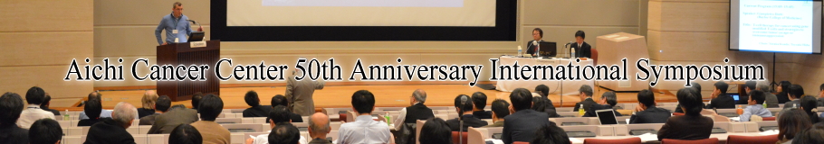
| Aichi Cancer Center 50th Anniversary International Symposium |
|
|
|
| Cancer Drug Resistance: Mechanisms and Strategies for Its Circumvention |
| 9:00〜17:50, March 14 (Sat.), 2015 |
International Conference Center, Aichi Cancer Center
1-1 Kanokoden, Chikusa-ku, Nagoya, Aichi 464-8681, Japan |
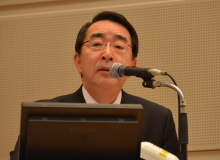
 Opening Remarks Opening Remarks |
 |
|
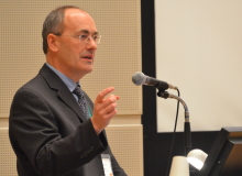
Taira Kinoshita
President, Aichi Cancer Center |
|
Good morning, distinguished guests, ladies and gentlemen.
On behalf of the organizing committee, It’s my pleasure and honor to have
an opening remarks on this special memorial international symposium to
celebrate the 50th anniversary of the Aichi Cancer Center.
Welcome to the symposium.
I would like to express my sincere gratitude and thanks to all the participants
especially to the invited speaker.
The Aichi Cancer Center was established in 1964 when we had Tokyo Olympic Games and the New Tokaido Line that is Shinkansen was opened in the same year.
In these fifty years Aichi Cancer Center worked very hard as a top runner
of the cancer clinics and the researches with the Cancer Institute and
the National Cancer Center.
The theme of the symposium is Cancer Drug Resistance---Mechanisms and Strategies
for it’s Circumvention, really a leading edge topic.
I would like to expect the fruitful hot discussions bring some new idea of the participants and will contribute to progress the cancer drug therapy itself.
Again welcome to the symposium and welcome to Nagoya.
Now it’s time to start the symposium.
Thank you. |
|
|
Opening Keynote Lecture
|
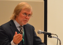
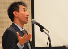
| カリフォルニア大学サンフランシスコ校のFrank McCormick先生に、「Targeting KRAS-induced stemness」のタイトルでご講演いただきました。 |
 |
|
Frank McCormick, Ph.D., F.R.S., D.Sc.
University of California, San Francisco (UCSF), USA
 Profile
 |
|
Targeting KRAS-induced stemness
Frank McCormick & Man-Tzu Wang
University of California, San Francisco - Helen Diller Family Comprehensive
Cancer Center 1450 3rd Street, Box 0128, San Francisco, CA 94158-9001,
USA
Of the three Ras genes, KRAS, NRAS and HRAS, KRAS is by far the major
contributor to human cancer, whereas HRAS is rarely activated. In spite
of this dramatic difference, KRAS and HRAS interact with the same effectors
and are equally potent at transforming cells in culture. However, cells
transformed by KRAS have unique properties relative to HRAS: they cause
a stem-like phenotype that enables them to grow as spheres in culture,
to establish tumors in mice at high efficiency and to resist the effects
of multiple chemotherapy and targeted drugs. These effects are due to KRAS’
ability to bind calmodulin, and to inhibit calmodulin-dependent kinase.
Low CaM kinase promotes wnt signaling and initiates a set of programs that
confer stemness. Binding of K-Ras to calmodulin is prevented by phosphorylation
of K-Ras on serine-181, by protein kinase C. Treatment of mice with a natural
product, prostratin, that activates PKC and K-Ras phosphorylation prevents
initiation of pancreatic tumors in xenograft models. Part of the “stemness”
program initiated by K-Ras involves secretion of the cytokine LIF, an IL-6
family member with a unique role in maintaining stemness. Neutralization
of LIF with a monoclonal antibody reduces stemness and sensitizes established
pancreas tumors to gemcitabine. We propose that attacking targets in these
stem-like pathways offers new opportunities for therapeutic intervention
in KRAS-driven cancers.
Key words: K-Ras, Stem cells, calmodulin, PKC |
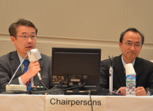
| Chair:Masahiro Aoki |
|
|
|
Session 1. Cancer Heterogeneity and Drug Resistance
|
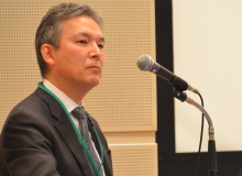
| 最初のセッションでは「がんの不均一性と薬剤耐性」をテーマに、3名の演者にご講演いただきました。 |
 |
|
Matthew J. Ellis, M.D., Ph.D.
Baylor College of Medicine, USA
Profile |
|
Genome-directed therapeutics for endocrine therapy resistant ER+ breast
cancer
Matthew J. Ellis
McNair and CPRIT Scholar, Director and Professor, Lester and Sue Smith
Breast Center, Baylor College of Medicine, Houston, TX, USA
As a result of improvements in DNA and RNA sequencing techniques the
genomic structure of estrogen receptor positive breast cancer is increasingly
well documented, but extracting clinically actionable information from
these complex data sets has proved fraught with difficulties. Barriers
to progress include the lack of pharmacological hypotheses for novel luminal
breast cancer tumor suppressor genes (e.g. MAP3K1, MLL3, SF3B1); 2) a lack
of a full understanding of interactions between mutation status, the prognosis
of ER+ breast cancer, and the effectiveness of endocrine therapy; 3) an
inadequate collection of patient-derived xenograft (PDX) models for luminal
breast cancer that fully encompass the heterogeneity of the disease; 4)
the logistical barriers of developing adjuvant strategies to exploit rare
drivers present in less than 5% of tumor samples; 5) insufficient genomic
discovery efforts directed towards samples accrued from patients suffering
from endocrine therapy resistant disease progression and 6) an incomplete
understanding of how complex somatic genotypes drive the biochemical events
responsible for the “hallmarks” of luminal cancer.
To better address these issues, five areas of investigation will be discussed: 1) somatic mutation diagnosis in DNA from primary breast cancer samples from patients treated with adjuvant tamoxifen and followed for over 20 years; 2) DNA and RNA sequencing of samples accrued from patients treated with neoadjuvant endocrine therapy to define the molecular origins of intrinsic aromatase inhibitor resistance and to identify pharmacological hypothesis; 3) efforts to expand and catalog patient-derived xenografts from ER+ breast cancers, including the use of mass spectrometry-based analysis of their proteomes and phosphoproteomes to expand our knowledge of the biochemistry of individual tumors; 4) a functional and pharmacological investigation of mutations in ESR1, including resistance-activating chromosomal translocations, and 5) the development of a neoadjuvant endocrine therapy strategy that identifies patients with intrinsic endocrine therapy resistance within a month of starting treatment so that they can be triaged to mutation-matched investigational treatment.
- Ellis MJ, and Perou CM. The genomic landscape of breast cancer as a
therapeutic roadmap.
Cancer Discov. 2013;3(1):27-34.
- Goncalves R, Warner WA, Luo J, and Ellis MJ. New concepts in breast
cancer genomics and genetics. Breast Cancer Res. 2014; 16(5): 460.
- Li S, et al. Endocrine-therapy-resistant ESR1 variants revealed by genomic
characterization of breast-cancer-derived xenografts. Cell Rep. 2013;4(6):1116-30.
- Tabchy A, Ma CX, Bose R, and Ellis MJ. Incorporating genomics into breast
cancer clinical trials and care. Clin. Cancer Res. 2013;19(23):6371-9.
|
 |
|
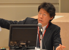
Tatsuhiro Shibata, M.D., Ph.D.
National Cancer Center, Tokyo, Japan
Profile |
|
Trans-ethnic landscape of hepatocellular carcinoma genomics
Tatsuhiro Shibata1, 2 , David A. Wheeler3, Hiroyuki Aburatani4
1Division of Cancer Genomics, National Cancer Center, Tokyo, Japan; 2Laboratory of Molecular Medicine, The Institute of Medical Science, 4Genome Science Division, Research Center for Advanced Science and Technology,
The University of Tokyo, Tokyo, Japan; 3Human Genome Sequencing Center, Baylor College of Medicine, Houston, TX,
USA
Multiple etiological factors (hepatitis virus infection, alcohol, obesity etc) are associated with the occurrence of hepatocellular carcinoma (HCC) and their contributions diverse among ethnicity. To elucidate genetic diversities in HCC genomes with regards to ethnic and epidemiological differences, we have conducted the trans-ethnic cancer genome research under the umbrella of the International Cancer Genome Consortium (ICGC) and The Cancer Genome Atlas (TCGA).
We performed whole exome sequencing of 514 pairs of HCC, which include
different ethnic populations (424 cases from the Japanese cohort and 90
from the US cohort) with various etiological backgrounds. Furthermore,
whole exome data of 105 HCC cases from TCGA was included in the mutation
signature analysis. Mutation call algorithms of three collaborating genome
centers (National Cancer Center, Tokyo, Research Center for Advanced Science
and Technology in the University of Tokyo, and Baylor College of Medicine,
Houston) were adjusted and validated by the Ion Proton sequencer. In total,
more than 100,000 somatic mutations were collected, and their signatures
were significantly associated with ethnicity and gender, but not with the
hepatitis virus status. In addition to TP53, WNT, and SWI/SNF pathways,
aberrant activation of the TERT pathway by various mechanisms (promoter/coding
mutations, gene amplification and viral genome integration) was found to
play a central role in hepatocarcinogenesis. Aggregation of the large cancer
genome data by ICGC and TCGA has rapidly progressed. In addition to the
cross-tumor analysis (Pan-Cancer study), population-based meta-cancer genome
analysis would provide us unique and diverse landscapes of the cancer genomes
on this planet. |
 |
|
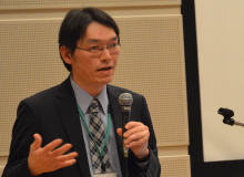
Tetsuya Mitsudomi, M.D., Ph.D.
Kinki University
Profile |
|
Acquired resistance in targeted therapy against driver gene mutation in
lung cancer
Tetsuya Mitsudomi1, Kenichi Suda1, Hiroshi Mizuuchi1, Yoshihisa Kobayashi1,Kazuto
Nishio2, Yasushi Yatabe3
Department of Thoracic Surgery1 and Genome Biology2, Kinki Unversity Faculty
of Medicine, Department of Pathology and Moleclular Diagnostics3, Aichi
Cancer Center Hospital, Japan
Discovery of activating mutation of the EGFR gene in adenocarcinoma of
the lung in 2004 opened the era of personalized therapy in thoracic oncology.
These tumors are highly dependent on the EGFR pathway and EGFR-tyrosine
kinase inhibitors (TKI) significantly prolong progression free survival
in these patients compared with chemotherapy. In 2007, EML4-ALK translocation
was found and these tumors are very sensitive to ALK-TKI. However, acquired
resistance inevitably develops usually after a median of 10 months. The
mechanisms for this resistance can be classified into 1) target gene alterations
(T790M mutation in EGFR-TKI or L1196M and other mutations in ALK, 2) activation
of additional kinases (e.g., MET, HER2 for EGFR, and KIT, EGFR, SRC for ALK) bypassing the inhibition
of the original kinases, and 3) other mechanisms including epithelial-mesenchymal
transition, small cell lung cancer transformation, etc.
To overcome T790M gatekeeper mutations, so-called third generation EGFR inhibitors that selectively inhibit EGFR-T790M while sparing the wild-type EGFR are being actively developed. Likewise, ALK-TKIs of a newer generation are active at least for some of the secondary mutations found in crizotinib-resistant tumors. Tumor resistance caused by the bypass track can be coped with by combination of the inhibitors for the original kinase and the bypassing kinases.
However, even with these strategies, cancer cells are smart enough to
escape from the therapy using other mechanisms. Heterogeneities in terms
of resistant mechanisms within a single patient become evident when specific
therapeutic pressure persists. Therefore, we also need to have armamentarium
that utilizes other mechanisms to cure lung cancer. Recent advances of
immunotherapy targeting PD-1/PD-L1 appear attractive in this respect. These
mechanism-driven therapeutic approaches will convert this fatal disease
into a more chronic disorder, and eventually into a curable disease with
the least patient burdens. |
| Chair: |
Takashi Takahashi |
|
Hiroji Iwata |
|
|
|
|
Session 2. Cancer stem cells, tumor dormancy, and drug resistance
|
| このセッションでは、「がん幹細胞、腫瘍の休眠状態と薬剤耐性」のテーマで、3名の先生にご講演いただきました。 |
 |
|
Nick Barker, Ph.D.
A*STAR Institute of Medical Biology, Singapore
Profile |
|
Lgr5+ stem cells in epithelial self-renewal and cancer of the stomach and
ovary
Nick Barker, Marc Leushacke, Annie Ng
A-STAR* Institute of Medical Biology, Singapore
The availability of robust cell-surface markers for identifying and isolating
adult stem cells is essential for studying both their normal in-vivo function
during tissue renewal and for evaluating their contribution to cancer.
Lgr5, a Wnt target gene expressing a 7-TM receptor that functions as facultative
component of the Wnt receptor complex, has been shown to selectively mark
stem cells in a range of rapidly renewing tissues, including the small
intestine, colon, stomach, hair follicle and developing kidney. Clonal
fate mapping employing the stem cell-specific Lgr5-CreERT2 line has been
used to further dissect how these adult stem cell pools maintain tissue
homeostasis and contribute to tissue repair following damage. Additionally,
targeted in-vivo mutation of the Lgr5+ve adult stem cell pools using the
same Lgr5-CreERT2 model has been used to determine the contribution of
stem cells to tumor initiation and progression in various epithelia. A
summary of the latest findings in the stomach and ovary will be presented
here. |
 |
|
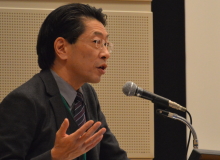
Hideyuki Saya, M.D., Ph.D.
Keio University
Profile |
|
Regulation of cell differentiation by actin dynamics and its application
in cancer treatment
Hideyuki Saya & Hiroyuki Nobuse
Division of Gene Regulation, Institute for Advanced Medical Research, School of Medicine,
Keio University, Tokyo, Japan
Differentiation status is strongly associated with the behavior of cancer
cells. Therefore, changes in the cellular context, which regulates the
differentiation potential, may serve in novel therapeutic strategies in
treating cancers.
We have established a mouse osteosarcoma (OS) model through overexpression
of c-MYC in bone marrow stromal cells (BMSCs) derived from Ink4a/Arf (-/-) mice. In this model, we found that the loss of adipogenic potential was
an essential event for OS development. Therefore, our understanding of
regulatory mechanisms of adipocyte differentiation would greatly contribute
to control OS tumorigenesis.
Adipocytic differentiation is accompanied by the adoption of a rounded
cell shape that is characteristic of mature adipocytes. Cell shape is determined
primarily by the actin cytoskeleton. We have recently found a novel regulatory
mechanism of adipocyte differentiation, in which regulation of transcriptional
coactivator MKL1 by actin cytoskelton dynamics drives adipocyte differentiation
mediated by PPARγ, a master transcriptional regulator of adipogenesis.
Accordingly, adipocyte differentiation can be induced by the disruption
of actin stress fibers through down-regulation of RhoA-ROCK signaling.
Based on this concept, we attempted to induce adipocyte differentiation
in OS cells, which resulted in a significant suppression of tumorigenesis.
Induction of trans-differentiation in cancer stem cells by regulating actin
cytoskeleton dynamics is a potential approach for some tumor types. |
 |
|
Masaki Inagaki, M.D., Ph.D.
Aichi Cancer Research Institute
Profile |
|
Cancer research on the two noteworthy issues: tetraploidy and primary cilia
Masaki Inagaki
Division of Biochemistry, Aichi Cancer Center Research Institute and Department
of Cellular Oncology, Nagoya University Graduate School of Medicine, Japan
Tetraploidy, a state in which cells have doubled chromosomal sets, is
observed in ~20% solid tumors and considered to frequently precede aneuploidy
in carcinogenesis. Tetraploidy is also detected during tissue differentiation
and aging process. We generated knock-in mice featuring vimentin with mitotic
phosphorylation-defective mutations to impair cytokinesis. Homozygotic
(VIMSA/SA) mice presented with microophthalmia and cataracts, in which lens epithelial
cells exhibited binucleation and aneuploidy, along with premature aging.
We further analyzed the ability to repair wounds in the skin of VIMSA/SA mice, and found that some subcutaneous tetraploid fibroblasts caused
by cytokinetic failure enter a new cell cycle and then develop into aneuploid
fibroblasts in vivo, which promotes premature aging. We suggest that tetraploidy without the
genetic alteration of cancer-related genes may be associated with premature
aging rather than carcinogenesis.
Non-motile primary cilia are microtubule-based sensory organelles that
regulate a number of signaling pathways during development and tissue homeostasis.
Tumor cells are known to often lack primary cilia, but whether their loss
is directly linked to tumorigenesis is completely unclear. We have recently
found that ubiquitin-proteasome machinery removes trichoplein, a negative
regulator of ciliogenesis, from mother centrioles and thereby causes Aurora-A
inactivation, leading to ciliogenesis. We have identified KCTD17 as a substrate-adaptor
for Cul3-RING E3 ligases (CRL3s) that polyubiquitylates trichoplein. Depletion
of KCTD17 specifically arrests ciliogenesis at the initial step of axoneme
(ciliary microtubule doublet) extension through aberrant trichoplein-Aurora-A
activity. We would like to discuss the relationship between primary cilia
and cancer stem cells, which may be implicated in drug resistance against
cancer chemotherapy. |
| Chair: |
Shinsuke Iida |
|
Yoshitaka Sekido |
|
|
|
|
Session 3. Strategies for circumvention of cancer drug resistance
|
| このセッションでは、「がんの薬剤耐性に対する克服戦略」をテーマに、3名の先生にご講演いただきました。 |
 |
|
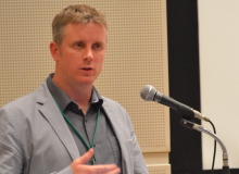
Gianpietro Dotti, M.D., Ph.D.
Baylor College of Medicine, USA
Profile |
|
T-cell therapy for cancer using gene modified T cells and strategies to overcome tumor escape or immunosuppression
Gianpietro Dotti
Center for Cell and Gene Therapy, Baylor Colledge of Medicine, Houston, TX, USA
T-lymphocyte-based treatments have enormous potential in cancer patients. Over the past decade, T cells mo dified to express chimeric antigen receptors (CARs) have had clinical success in B-lymphocyte derived malignancies. In the specific context of CAR-T cells therapies for B-cell malignancies we developed at Baylor a strategy aimed at achieving antitumor effects, but limiting the prolonged B-cell aplasia caused the infusion of CD19-CAR-specific T cells. We are currently targeting the k-light chain of human immunoglobulins expressed on the cell surface of k+lymphoma cells in an effort to target lymphomas cells but spare normal l+B-lymphocytes. An update of the clinical trial currently ongoing will be presented.
In contrast to B-cell malignancies, the clinical efficacy of CAR-T cells
remains limited in solid tumors. This unfavorable outcome could be due
to the insufficient migration of the infused T cells to the tumor site
and to the immunosuppressive characteristics of the tumor environment,
which inhibit the effector function and proliferation of those few T cells
that do reach the tumor. We recently found that tumor-specific engineered
T lymphocytes expanded ex vivo for adoptive T-cell therapy are defective in their capacity to degrade
one critical component of the extracellular matrix. We also found that
this defect can be however repaired by the ectopic expression of the enzyme
heparanase. We also found that armed oncolytic viruses expressing RANTES
and IL-15 can be used to favor the migration of CAR-T cells at the tumor
site and promote the survival of CAR-T cells within the hostile tumor environment. |
 |
|
Masaaki Komatsu, Ph.D.
Niigata University
Profile |
|
Loss of autophagy causes metabolic changes through a transcription-factor
pathway
Masaaki Komatsu
Department of Biochemistry, School of Medicine, Niigata University, Japan
Autophagy provides starved cells with amino acids, free fatty acids,
and glucose for new protein synthesis energy production; autophagy also
controls the quality and quantity of organelles such as mitochondria. Therefore,
it is plausible that autophagy might be integrated with metabolic pathways.
Indeed, suppression of autophagy causes myopathy, tumorigenesis, and metabolic
disorders in mice and humans. However, the metabolic changes associated
with deficiencies in autophagy are largely unknown. Furthermore, it remains
unclear whether the major predisposing factor for the aforementioned diseases
in the absence of normal autophagic activity is a simple deficit in supply
of molecular building blocks, dysregulation of mitochondrial homeostasis,
or some other cause. Here, we show that deficiencies in autophagy are associated
with rearrangement of glucose and glutamine metabolism via a transcriptional
regulatory mechanism. |
 |
|
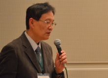
Yasuaki Arai, M.D., Ph. D.
National Cancer Center Hospital, Tokyo, Japan
Profile |
|
Interventional radiology in oncology
Yasuaki Arai
National Cancer Center Hospital, Tokyo, Japan
IR is a minimally invasive treatment modality in which small devices
are percutaneously inserted into a patient’s body with minimum incision
under image guidance.
There are two routes to access to the target lesion; trans-canal and
direct puncture. The typical type of trans-canal approach is transarterial
chemoembolization (TACE) for hepatocellular carcinoma (HCC), in which the
feeding arteries are occluded with anticancer drug to kill tumor cells
with stasis of blood flow. TACE could obtain total necrosis if the HCC
tumor is hyper-vascular and less than 5cm in diameter. In a decade, microspheres
with drug eluting and Yttrium-90 have been developed to treat HCCs with
various stages. The other approach with percutaneous direct puncture is
thermal ablation, such as radiofrequency ablation (RFA), microwave ablation,
cryoablation for tumors in the liver, kidney, lung, etc. TACE and RFA are
established as the standard treatment for early and intermediate stage
HCC.
Moreover, there are novel IR treatments; high-intensity focused ultrasounds (HIFU) and irreversible electroporation (IRE). HIFU kills tumor cells with thermal ablation by high-intensity focused ultrasounds without needle puncture. IRE kills tumor cells with membrane with electroporation by high voltage pulse without the destruction of anatomical structures.
IR can be complementary with other treatment modalities because the mechanism of anti-tumor effect in IR is completely different from that of medical and radiation therapy.
On the other hand, IR is difficult to establish evidence by clinical
trials, because the clinical results in IR greatly depend on the operator’s
skills and equipment. We started to conduct many multi-institutional clinical
trials in Japan more earlier than western countries, however, still it
is very challenging for us to establish IR as one of the standard treatments
in the oncology field. |
| Chair: |
Yutaka Kondo
|
|
Toyoaki Hida |
|
|
|
|
Closing Keynote Lecture
|
| 京都大学の長田重一先生に、「Apoptosis and exposure of phosphatidylserine」のタイトルでご講演いただきました。 |
 |
|
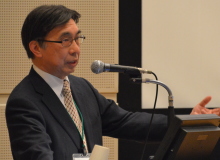
Shigekazu Nagata, Ph.D
Kyoto University
Profile |
|
Apoptosis and exposure of phosphatidylserine
Shigekazu Nagata
Department of Medical Chemistry, Graduate School of Medicine, Kyoto University, Japan
Apoptotic cells are swiftly engulfed by macrophages. If this process
does not occur properly, materials released from dead cells activate the
immune system, leading to systemic lupus erythematosus-type autoimmune
disease. Phospholipids in plasma membranes are asymmetrically distributed
between inner and outer leaflets, and phosphatidylserine (PtdSer) is exclusively
localized in the inner leaflet. The asymmetrical distribution of phospholipids
is maintained by an ATP-dependent phospholipid translocase or flippase.
When cells undergo apoptosis, or platelets are activated, the asymmetrical
distribution of phospholipids is disrupted by scramblase, leading to PtdSer-exposure.
The PtdSer exposed on dead cell surface is recognized by macrophages as
an “eat me” signal, while PtdSer on activated platelets provides the
scaffold for clotting factors. We recently identified two membrane proteins
(TMEM16F and Xkr8) as phospholipid scramblases, and a pair of membrane
proteins (ATP11C and CDC50A) as a flippase. TMEM16F, a protein with 8 transmembrane
regions, requires Ca2+ to support phospholipid scrambling, and plays an
essential role in the PtdSer-exposure in activated platelets. Xkr8 is a
protein carrying 6 transmembrane regions, and caspases cleave off its C-terminal
tail to promote the scramblase activity. ATP11C is a P4-type ATPase at
plasma membrane, and CDC50A works as a chaperone to transport ATO11C from
endoplasmic reticulum to plasma membranes. ATP11C translocates PtdSer from
outer to inner leaflets of plasma membranes in an ATP-dependent manner.
When cells undergo apoptosis, ATP11C is inactivated by caspase-mediated
cleavage, indicating that in addition to the caspase-mediated activation
of scramblase, inactivation of flippase is required to expose PtdSer during
apoptosis. Lymphoma cells that lack the flippase constitutively expose
PtdSer, are engulfed by macrophages, and can not develop tumors in nude
mice. These results indicate that PtdSer is necessary and sufficient as
an “eat me” signal to be recognized by macrophages, and the PtdSer-expressing
tumor cells can be killed by being engulfed by macrophages. |
| Chair: |
Tomohiro Kinoshita
|
|
|
|
|
Closing Remarks
|
 |
|
Masahiro Aoki
Aichi Cancer Center |
|
On behalf of the organizing committee, I would like to express our sincere gratitude to all the participants of the Symposium for making this symposium fruitful and successful.
Resistance to chemotherapy and targeted therapies, intrinsic or acquired, is clearly one of the major and most difficult problems we cancer researchers, basic or clinical or translational, are facing today and will most likely be facing in the near future as well, and that's why we chose the topic as the theme for this memorial symposium.
Today, we've learned so much about various aspects of the paradigm of cancer drug resistance, from the underlying basic biology to novel strategies for circumventing or overcoming resistance, aimed at better clinical management in future.
All the talks were absolutely wonderful, and I would like to thank our distinguished speakers again for kindly accepting our invitation and coming all the way to Nagoya, from abroad and from all over Japan despite their busy schedule. I also thank session chairs for providing an open and encouraging atmosphere for discussion. And of course, we are so grateful to you, the audience, numbering more than 200, from Hokkaido to Kyushu. Thank you so much for coming and actively participating in the symposium throughout the day.
As our president Dr. Kinoshita mentioned in his opening remarks, Aichi Cancer Center has a long and glorious history of 50 years as the third oldest comprehensive cancer center in Japan. It is the hope of the organizing committee and all the current members of Aichi Cancer Center that we will live up to the fame achieved by hard work of former members and to the expectations of the society. We also hope this symposium will help to develop further collaborative efforts in future.
Now, please allow me to take this opportunity to express may thanks to the members of the organizing committee, a mixed team from Hospital, Research Institute, and Administration Office, as well as to our Cancer Center staffs who played various roles today for making this symposium possible.
Finally, for those of you who are travelling back home, I wish you a safe and pleasant trip.
Thank you all again, and see you in 50 years! |
50周年記念国際シンポジウム実行委員会
Organizing Committee
青木正博(委員長)
Masahiro Aoki (Chairperson)
岩田広治(副委員長)
Hiroji Iwata (Vice-Chairperson)
樋田豊明
Toyoaki Hida
木下朝博
Tomohiro Kinoshita
室 圭
Kei Muro
小島 康
Yasushi Kojima
細野覚代
Satoyo Hosono
笠原広介
Kousuke Kasahara
関戸好孝(アドバイザー)
Yoshitaka Sekido (Advisor) |
|
|